How is Mesothelioma Diagnosed? | X-ray, CT Scan, MRI Scan, PET Scan
If you are showing symptoms of mesothelioma, a medical Doctor will look at your medical history, provide a medical exam, and review a number of diagnostic tests in order to confirm or rule out mesothelioma. The medical provider will need to carefully review your history of medical treatment as well as evaluate risk factors and evaluate particular symptoms.
How is Mesothelioma Diagnosed?
Mesothelioma is a rare and aggressive form of cancer that affects the mesothelial lining of various organs, most commonly the lungs. Diagnosing mesothelioma typically involves a combination of medical history assessment, physical examination, and various imaging and diagnostic tests. The tests commonly used in the diagnostic process include X-rays, CT scans, MRI scans, and PET scans, among others. Here’s an overview of how mesothelioma is diagnosed:
- Medical History and Physical Examination: A thorough medical history is taken to identify potential risk factors such as asbestos exposure. A physical examination may reveal signs and symptoms of mesothelioma, such as pleural effusion (fluid in the lungs) or abdominal swelling (ascites).
- Imaging Studies:
- Chest X-ray: A chest X-ray is often one of the initial diagnostic tests. It can reveal pleural effusion or other abnormalities in the lungs.
- CT Scan (Computed Tomography): CT scans provide detailed cross-sectional images of the chest or abdomen. They can help identify the presence and extent of tumors, as well as any spread to nearby structures.
- MRI Scan (Magnetic Resonance Imaging): MRI scans can offer additional information, especially when evaluating the extent of mesothelioma in the chest or abdomen. They can provide better soft tissue contrast than X-rays or CT scans.
- PET Scan (Positron Emission Tomography): PET scans are used to identify areas with increased metabolic activity, which can indicate the presence of cancer. They are often used in combination with CT scans to provide more precise information about the extent of the disease.
- Biopsy: To confirm the diagnosis of mesothelioma, a biopsy is usually required. A biopsy involves taking a sample of tissue from the affected area for examination under a microscope. There are different types of biopsies, including:
- Needle Biopsy: A thin, hollow needle is used to extract tissue samples.
- Thoracoscopy or Laparoscopy: These minimally invasive procedures involve the insertion of a thin tube with a camera into the chest or abdomen to visualize and collect tissue samples.
- Open Surgical Biopsy: In some cases, open surgery may be required to obtain tissue samples for a biopsy.
- Laboratory Tests: The tissue samples obtained through the biopsy are examined by a pathologist to determine the presence of mesothelioma cells. Specialized tests, such as immunohistochemistry, can help differentiate mesothelioma from other types of cancer.
- Staging: Once mesothelioma is confirmed, further imaging tests, such as additional CT or MRI scans, may be performed to determine the stage of the cancer and assess its spread. This information is crucial for developing a treatment plan.
The combination of these diagnostic methods helps determine the presence of mesothelioma, its type (e.g., pleural, peritoneal, pericardial), its stage, and the extent of the disease. Early diagnosis is important for improving the chances of successful treatment. If you or someone you know has been exposed to asbestos or is experiencing symptoms associated with mesothelioma, it’s essential to consult a healthcare professional for proper evaluation and diagnosis.
Mesothelioma is a rare and aggressive form of cancer that primarily affects the mesothelium, the thin membrane lining the chest and abdominal cavities. Diagnosing mesothelioma typically involves a combination of medical history evaluation, physical examinations, and various imaging and diagnostic tests. Here are some of the common methods used to diagnose mesothelioma:
- Medical History and Physical Examination: The diagnostic process often begins with a detailed medical history, where the patient discusses their exposure to asbestos, which is a known risk factor for mesothelioma. A physical examination is also performed to check for any signs and symptoms of the disease.
- Imaging Tests:
- X-ray: A chest X-ray is often the initial imaging test used to detect abnormalities in the lungs and chest cavity.
- CT Scan (Computed Tomography): A CT scan provides more detailed cross-sectional images of the chest and abdomen, helping to visualize the extent of the tumor and its potential spread.
- MRI Scan (Magnetic Resonance Imaging): An MRI can offer better soft tissue detail and help in assessing the tumor’s size, location, and involvement of nearby structures.
- PET Scan (Positron Emission Tomography): PET scans use a radioactive tracer to detect areas of increased metabolic activity. Mesothelioma cells often have increased metabolism, making PET scans useful for staging and determining the extent of the disease.
- Biopsy: A biopsy is the definitive method for diagnosing mesothelioma. During a biopsy, a small tissue sample is collected from the suspicious area (usually the pleura, peritoneum, or pericardium) and examined under a microscope. There are different types of biopsies:
- Fine Needle Aspiration (FNA): A thin needle is used to extract a small sample of tissue or fluid.
- Core Needle Biopsy: A larger needle is used to remove a more substantial tissue sample.
- Surgical Biopsy: In some cases, a surgeon may need to perform a more invasive procedure, such as a thoracoscopy (for pleural mesothelioma) or laparoscopy (for peritoneal mesothelioma), to obtain tissue samples.
- Laboratory Tests: Once a tissue sample is obtained, it may be subjected to various laboratory tests, including immunohistochemistry, which helps identify the specific type of mesothelioma (e.g., epithelioid, sarcomatoid, or biphasic).
- Blood Tests: Some blood markers, such as mesothelin and soluble mesothelin-related peptide (SMRP), may be elevated in mesothelioma patients. However, these markers are not definitive for diagnosis and are often used in conjunction with other diagnostic methods.
The combination of these diagnostic methods allows healthcare professionals to accurately diagnose mesothelioma, determine its stage, and develop a treatment plan tailored to the patient’s specific situation. It is essential to consult with a healthcare specialist if you suspect you may have mesothelioma or have a history of asbestos exposure, as early detection and diagnosis can significantly impact treatment outcomes.
Physical Exam and health history
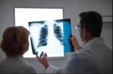
The medical provider will be interested in knowing whether you have been exposed to asbestos. The inquiry from the medical provider will look into 3 main factors:
- Where exposure to asbestos could have occurred in your past employment or history
- The duration of asbestos exposure
- Amount of exposure to asbestos
Signs of Mesothelioma
The medical provider will be looking for symptoms of mesothelioma such as:
- Pleural Effusion
- Ascites
- Pericardial effusion
- Shortness of breath
- Pain
What diagnostic tests will be used to determine if I have mesothelioma?
Medical providers utilize a variety of diagnostic image tests to determine whether there is Cancer. These diagnostic tests also evaluate how cancer may have progressed. The key part of the diagnostic test is to provide information to the medical provider to help make a diagnosis. The most used types of diagnostic testing include:
- X-Ray
- CT Scan
- MRI Scan
- PET Scan
- Biopsies
- Needle Biopsy
- Surgical Biopsy
How is Mesothelioma Diagnosed?
X-Ray- “X-rays are a type of radiation called electromagnetic waves. X-ray imaging creates pictures of the inside of your body. The images show the parts of your body in different shades of black and white. This is because different tissues absorb different amounts of radiation.” Medline
CT Scan- “A computerized tomography (CT) scan combines a series of X-ray images taken from different angles around your body and uses computer processing to create cross-sectional images (slices) of the bones, blood vessels and soft tissues inside your body. CT scan images provide more-detailed information than plain X-rays do.” Mayo Clinic
MRI Scan- “Magnetic resonance imaging (MRI) is a medical imaging technique that uses a magnetic field and computer-generated radio waves to create detailed images of the organs and tissues in your body. Most MRI machines are large, tube-shaped magnets. When you lie inside an MRI machine, the magnetic field temporarily realigns water molecules in your body. Radio waves cause these aligned atoms to produce faint signals, which are used to create cross-sectional MRI images — like slices in a loaf of bread” Mayo Clinic
PET Scans- “A positron emission tomography (PET) scan is an imaging test that allows your doctor to check for diseases in your body. The scan uses a special dye containing radioactive tracers. These tracers are either swallowed, inhaled, or injected into a vein in your arm depending on what part of the body is being examined. Certain organs and tissues then absorb the tracer.” Healthline
Biopsies:
Biopsies are a crucial diagnostic procedure widely utilized by medical providers for suspected victims of asbestos exposure especially victims with signs any symptoms of mesothelioma. Biopsies are lee invasive than surgical biopsies and surgery.
“A biopsy is a sample of tissue taken from the body in order to examine it more closely. A doctor should recommend a biopsy when an initial test suggests an area of tissue in the body isn’t normal. Doctors may call an area of abnormal tissue a lesion, a tumor, or a mass. These are general words used to emphasize the unknown nature of the tissue. The suspicious area may be noticed during a physical examination or internally on an imaging test.” Web MD
Needle Biopsy- “A needle biopsy is a procedure to obtain a sample of cells from your body for laboratory testing. Common needle biopsy procedures include fine-needle aspiration and core needle biopsy. Needle biopsy may be used to take tissue or fluid samples from muscles, bones, and other organs, such as the liver or lungs.” Mayo Clinic
Surgical Biopsy- A surgical Biopsy is an open surgery. It is invasive compared to a regular biopsy. A Surgical Biopsy is usually done when other biopsy results are inconclusive. “A procedure in which a cut is made through the skin to remove abnormal tissue so it can be checked under a microscope for signs of disease. The two types of surgical biopsy are incisional biopsy, in which part of a lump or a sample of tissue is removed, and excisional biopsy, in which an entire lump or suspicious area is removed. Also called open biopsy.” Cancer.gov
Pleural Biopsy- “A biopsy is a procedure done to remove a tissue sample from the body so it can be examined. The pleura is a double layer of membranes that surrounds the lungs. A pleural biopsy is a procedure to take a small piece of the pleura. This is done with a special biopsy needle. Or it’s done during surgery. The biopsy is done to look for infection, cancer, or another condition.” Hopkins Medicine
Thoracentesis- “Thoracentesis is a procedure to remove fluid or air from around the lungs. A needle is put through the chest wall into the pleural space. The pleural space is the thin gap between the pleura of the lung and of the inner chest wall.” Hopkins medicine
What Is a Thoracotomy?
“A thoracotomy is a surgical procedure in which a cut is made between the ribs to see and reach the lungs or other organs in the chest or thorax. Typically, a thoracotomy is performed on the right or left side of the chest. An incision on the front of the chest through the breast bone can also be used, but is rare. A thoracotomy is performed for diagnosis or treatment of a disease and allows doctors to visualize, biopsy or remove tissue as needed.” Lung.org
What is a Thoracoscopy?
“Thoracoscopy is a procedure a doctor uses to look at the space inside the chest (outside of the lungs). This is done with a thoracoscope, a thin, flexible tube with a light and a small video camera on the end. The tube is put in through a small cut made near the lower end of the shoulder blade between the ribs. Thoracoscopy is sometimes done as part of a VATS procedure, which is short for video assisted thoracic surgery.” Cancer.org
What is a Laparotomy?
This type of surgery is very similar to a Thoracoscopy but the incision in this surgery is in the abdomen not near the lower end of the shoulder blade as in a Thoracoscopy. “A laparotomy is a surgical incision (cut) into the abdominal cavity. This operation is performed to examine the abdominal organs and aid diagnosis of any problems, including abdominal pain. In many cases, the problem – once identified – can be fixed during the laparotomy. In other cases, a second operation is required. Another name for laparotomy is abdominal exploration.” Better health
What is a Laparoscopy?
“Laparoscopy is a type of surgical procedure that allows a surgeon to access the inside of the abdomen (tummy) and pelvis without having to make large incisions in the skin. This procedure is also known as keyhole surgery or minimally invasive surgery.” nhs
What is a Mediastinoscopy?
Surgeons use this procedure to look at the lymph nodes and collect samples to see if cancerous. This allows surgeons to determine whether cancer has spread or is localized. This procuder will help surgeons differntiate mesothelioma from lung cancer. This biopsy will help asses whether other surgery is needed to remove cancer. “A procedure in which a mediastinoscope is used to examine the organs in the area between the lungs and nearby lymph nodes. A mediastinoscope is a thin, tube-like instrument with a light and a lens for viewing. It may also have a tool to remove tissue to be checked under a microscope for signs of disease. The mediastinoscope is inserted into the chest through an incision above the breastbone. This procedure is usually done to get a tissue sample from the lymph nodes on the right side of the chest.” Cancer.org
If you have developed cancer or mesothelioma as a result of asbestos exposure, contact us for a free review to determine if you can file a mesothelioma lawsuit.